


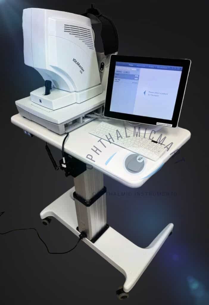
Nidek RS-330 Retina Scan Duo is a combined OCT and fundus camera system that is a user-friendly, versatile unit that provides high-definition images and value-added features.
For user-friendly OCT, the Nidek RS-330 Retina Scan Duo is a piece of ideal equipment for your practice. It is very easy to operate and does not require high technical skills. The Nidek RS-330 retina scan duo will guarantee high-quality output and efficient operations, making it suitable for use across practices of different sizes.
The boost in productivity and efficiency that this model will guarantee is only part of its great appeal. But what are other features of the Nidek RS-330 that are tailored to suit the needs of your practice?
The intuitive software, the automated functions, the rapid measurements, and high-quality images make the Retina Scan Duo a pleasure to operate, akin to photography that captures many of the vivid landscapes experienced over your lifetime. The combination of features results in a better overall experience for the patient and practitioner.
Features and benefits of the Nidek RS-330 Retina Scan Duo.
Auto tracking
Nidek’s acclaimed 3D auto-tracking and automatic shot allow for a user-friendly imaging process of the fundus and all its properties. Both the professional and standard modes have a customizable interface, allowing you to make a choice based on your clinic’s preferences. This automatic tracking and shooting make it easy to delegate functions to different clinicians because the process requires minimal operator interference.
High-quality imaging
 The brand’s commitment to high-quality imaging is maintained by the Nidek RS-300 retina scan duo. This OCT will average a large number of very high-quality images, allowing your operator to determine a diagnosis better.
The high-quality images can be used for patient tracking, allowing you to monitor the progress of any treatment plans you may have instituted. This will enable you to offer comprehensive services to your patients instead of referring them to other practices.
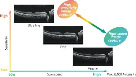
For OCT imaging, up to 50 images can be averaged and the OCT sensitivity is selectable among ultra-fine, fine, and regular sensitivities based on ocular pathology. The Retina Scan Duo™ has a built-in 12-megapixel CCD camera, producing high-quality fundus images.
Large area scan
The wide area macula centered scan is the key image of the Nidek RS-330 Retina Scan Duo. This equipment provides a color-coded guide map to compare the patient’s macular thickness with the expected standard. Your practitioner will be better able to make diagnosis thanks to the comprehensive layout and comparison by the large scan.
Multiple patterns
You are not limited to any one type of scan. This module allows for a variety of OCT scanning patterns for different ocular and retinal regions for better visualization. It can also accommodate an anterior adapter to help display multiple patterns on this part of the eye as well. Your operator will be able to scan the eye comprehensively and make a more accurate diagnosis.
Value-added features
Value-added features will improve your experience with the Nidek RS-330 Retina Scan Duo. You can install additional features through En Face OCT and fundus autofluorescence (FAF) modules. This will increase the variety of options available to your clinician when making the final call. However, some features may be exclusive to specific models.
Multiple reports
Aside from multiple patterns, the Nidek RS-330 Retina Scan Duo can present different reports to help an operator make a better overall diagnosis. It can present a report for both eyes, as well as the option of a macula map or line. The more details provided about the eye situation, the easier it will become for the clinician to make a great call in terms of both diagnosis and treatment suggestions.
OCT image screening software
The optional OCT image screening software* categorizes OCT images acquired from all models in the RS series, increasing efficiency when reviewing numerous OCT images.
*Analysis is performed with a software module created by CRESCO Ltd.
If the analysis outcome is blank, screening has not been performed or the images are not appropriate for screening.
Screening Condition
1) Macula cross, Macula multi cross, Macula radial 6/12 and Macula map (only if a cross scan is executed) scan pattern
2) Scan length is 6.0 mm 3) Scan position is 0 or 90 degrees 4) Image centered on the fovea
FEATURES
- User-Friendly Interfaces for Two Capture Modes
- 3-D Auto Tracking and Auto Shot
- Operation with Joystick for Flexible Alignment
- Space-saving Unit
- HD Image Averaging (max. 50 images)
- Selectable OCT Sensitivity – ultra-fine, fine, regular
- Enhanced Image
- Wide Area Scan (12 x 9 mm) / Wide Area Normative Database
- Multiple OCT Scan Patterns
- 12-megapixel CCD Camera
- Stereo and Panorama Photography
- Fundus Autofluorescence (FAF)*1
- En face OCT
- NAVIS-EX
- Anterior Segment Adapter*4
Reference Library
Nidek RS-330 Retina Scan Duo Manufacturer Information
Nidek RS-330 Retina Scan Duo Brochure


Reviews
There are no reviews yet.