
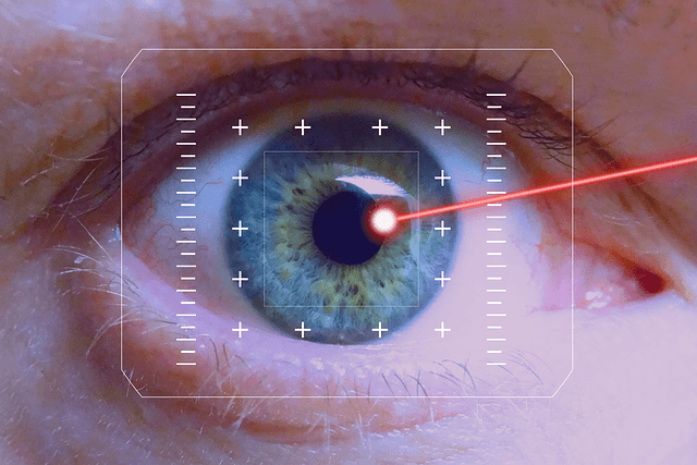
Corneal Topography Machines are a technological procedure designed to monitor and measure changes that might occur in the shape of the eye’s cornea. It can also be defined as the diagnostic tool assisted by the computer to create a three-dimensional figure of the curvature surface of the eye’s cornea.
Have you ever been diagnosed with an eye complication? Nearly everyone has been the victim of any single-eye implication at some point. However, there is a more significant issue here.
Interestingly, you cannot successfully diagnose any eye disease; why? This is because the doctor cannot see everything happening in the eye. However, this can only be done by using a corneal topography machine to help make the diagnosis.
What is Corneal Topography?
Have you ever been involved in any contact lens examination? If you have ever been in such a situation, you have witnessed the doctor charging an extra fee for corneal topography.
Then, what optical corneal topography are you being charged extra cash for? I will provide you with detailed information on what it is all about. So, if you are in the same situation next time, I want you to know what the doctor is charging you extra cash.
The eye’s cornea is responsible for almost about seventy percent of the eye’s focusing power. This is because normal sight vision typically has an evenly rounded cornea.
However, if the cornea is too flat, steep, or has an unevenly curved surface, there will be a result of a vision that could be better. Thus, the doctor will charge you extra cash and take advantage of Corneal Topography Machines, as they are suitable for detecting such changes if they are in your eye. Thus, this is the most convenient test to be done.
Knowing the corneal topography, what is this cornea we are emphasizing? It is good to know what it is so that you have a better understanding of identifying the defects that it might possess. Therefore, here is what cornea is as far as optometry is concerned.
It is a transparent dome-shaped tissue located in the eye, and it’s majorly known to cover the iris and the pupil. Besides, it provides about two-thirds of the refracting power to an individual’s vision. It is made up of specialized cells, thus making it a more remarkable tissue.
However, it doesn’t have any blood vessels in it that can provide it with nourishment. Therefore, this makes it receive most of its food directly through the aqueous humor from the eye and the tears on the surface. However, it is not a defect if the cornea has no blood vessels. This is because it is like a lens; thus, it must be transparent without any blood vessels that might interfere with eye focusing.
How does Optical Corneal Topography Machines Work?

Knowing what corneal topography is, it is also advisable to understand how it works. So, I will explain how the system works to ensure the topographer gets the full details from the cornea.
The optical corneal topography machines produce a detailed, visual description of the corneal shape and power. By working to provide this analysis, the doctor will get excellent details about the cornea conditions and the corneal surface.
All these achievements are after a series of illuminated rings known as the calm disc projected on the cornea surface. These reflections are in the instrument.
The computer then generates a topographical map of the cornea after the analysis of the mirrored rings has been done. After this, the details provided by the network can be helpful in the diagnosis. This included monitoring and the treatment of various conditions present in the eye. Besides, they can be used in contact lens fitting and surgery planning, including laser vision correction.
During diagnosis, the patient sits in front of the bowl, and the head is pressed against a bar when data points are generated. This is because the optical corneal topography machines have a computer that links to a lighted bowl with a pattern of rings.
Computer software then digitizes these data points to produce a printout of the corneal shape. Usually, To identify different elevations, different colors are applied, like how the topographic map of the earth displays is always available on the land surface.
Knowing how corneal topography works is a big plus, but the question is, what conditions are shown on the computer? You might have been asking yourself what they are, but you don’t have to worry, and I will take you through the conditions shown in detail. Therefore, below is what the optical corneal topography reveals during diagnosis.
Corneal topography machines are beneficial because they provide a wealth of information to the eye in different areas as follows:
- Keratometry: this older technology was used before the corneal topography was developed. It measures only a small surface area of the cornea, thus giving the doctor only two measurements: the cornea’s steepness. However, optical corneal topography technology has revolutionized corneal shape analysis. Therefore, instead of measuring only two points, it provides hundreds of data points of the cornea.
This helps build a correct mapped color of the entire steepness of the eye in either location the doctor is interested in analyzing. The technology, therefore, reveals to the doctor how much astigmatism you have. This is by generating a color map showing the steeper areas in reading and flatter areas in blue.
- Elevation maps: The technology also reveals the elevation maps to the doctor. This will help the doctor see the unusual spots on the cornea that are not the same. Besides, some software has also been developed to provide three-dimensional images that doctors can rotate in any direction they are interested in. This is to understand better what might be occurring in the cornea.
- Contact lens fitting: your doctor needs to watch you detect the changes in your eyes to avoid some severe complications. Therefore, the optical corneal topography comes in because it allows the doctor to determine the exact lens that can fit you. This is by revealing the precise shape of your cornea.
- Dry eye evaluation: the corneal topography reveals precisely how the surface of your eye is rough and, at the same time, how your vision is affected by a dry eye. This is because the tears in the eyes are very complex; thus, it cannot be straightforward to identify eye complications directly using a microscope.
Optical Corneal Topography Benefits
Corneal topography is not a routine test as most people think; however, its application is in diagnosing specific implications.
Apart from this, its form evaluates disease progression, contact lens fitting, and surgery planning. However, the technology is commonly employed in refractive eye surgeries.
Besides, the map produced by the device is used together with other tests to determine how much corneal tissue can be removed to correct the visual defect.
In the diagnosis and management use of the optical corneal topography, specific abnormalities and diseases that are dealt with are not limited to the following:
- Corneal transplants
- Corneal opacities
- Corneal deformities
- Fitting contact lenses
- Planning for refractive surgeries
- Irregular astigmatism following the corneal transplantation.
Knowing the uses of corneal topography is also good, as are its benefits. Therefore, below are its benefits that you may need to know:
Here are the benefits of an optical corneal topography that you may need to know
- It retains patients
- Specialty lens wearers usually produce more substantial profits
- The specialty wears are very loyal
- Several specialty lenses candidates exist
The Latest Corneal Topography Machines in Ophthalmology
Health and technology are directly related and depend on each other. Why am I saying this? When it comes to health problems, especially those associated with the eye, you need assistance to make some treatments. The vision is a very delicate structure among the body organs; thus, it cannot just be approached like other organs.
As a result, scientists have developed many devices that have helped diagnose and manage the implications in the eye. To add, they have been working hard on improving these devices daily to ensure that all details or any changes on the eye are provided.
As a result, optical corneal topography is one of the tools used for decades in eye problem diagnosis. This device has been of benefit to doctors who specialize in eye treatments. Because of this, some corneal topographers have emerged in ophthalmology.
Here are 10 of the latest optical Corneal Topography Machines in ophthalmology.
1. Oculus easygraph ($2,999.00)
Oculus easygraph offers the ideal solution for those practices that struggle with space limitations. It, therefore, incorporates the cornea assessment into the examination process by mounting directly on the slit lamp.
Oculus easygraph design uses the same precise measuring technology as that of keratograph. However, it is both a topographer and a keratometer that has been combined to become a unit. Besides, it also has numerous analyses and formats to support the cornea’s quantitative classification.
Contact fitting lenses can also be integrated using an Oculus easy graph to add confidence and enhance trust among contact lens consultants.
2. Galilei G4 ($9,999.00)

Galilei g4 integrates placid disc topography with the scheimflug topography as a single device. It allows for analyzing both the anterior and posterior surfaces of the corneal.
Galilei G4 concurrently uses two cameras for both different and pace testing approaches. Placido provides anterior surface irregularity detection, anterior corneal curvature, and tear film quality analysis.
This makes the recorded dual-scheimflug images produce reliable pachymetry with posterior curvature data. However, the calm generates high-quality central corneal curvature data fitted to the anterior corneal surface.
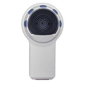
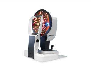
3. Cassini corneal ($5,900.00)
Cassini corneal helps cataract surgeons to understand corneal properties better. Thus, this helps improve outcomes and increase premium patient volume.
Cassini is a first-of-its-kind anterior segment ocular device used in diagnosis based on color-led technology. Cassini Total Corneal Astigmatism helps surgeons understand properties to improve outcomes and increase individual premium volume. Cassini is a first-of-its-kind anterior segment ocular device based on Colour LED Technology.
Besides, it also enables definitive diagnosis for corneal astigmatism, analysis for dry eye, contact lens fitting, planning for refractive cataract surgery, and corneal transplant.
4. Topcon ca-200f ($3,999.00)

Topcon ca-200f is a reasonably based topography system designed to deliver accurate images of the anterior corneal surface with a higher resolution. The twenty-four rings on it measure up to about ten thousand data points.
Topcon ca-200f also incorporates eight blue LEDs required for fluorescein images. These are essential for the simulation of contact lenses. The advantage of it is that it can be operated as a single stand unit or in combination with other external PCs.
To make the operation and evaluation of the cornea fast and easy, you can use a ten-inch touch screen with a high-speed CCD camera. This has auto-capture and the best features of an image.
5. Orbscan 3 ($5,555.00)
The Orbscan 3 is from Bausch + Lomb as an anterior segment analyzer and the third generation of the multidimensional orbscan topographer. It is a new platform with several features that have many enhancements driven by more than twenty years of use in the field.
Orbscan 3 is an indispensable screening tool to increase the number of ophthalmic surgeons with new refractive guidelines.
Orbscan 3 allows surgeons to examine optical pachymetry and anterior and posterior astigmatism, providing information points on equilibrium and biomechanics to inform choices and identify suitable candidates for refractive procedures.
6. Tracey iTrace ($3,999.00)
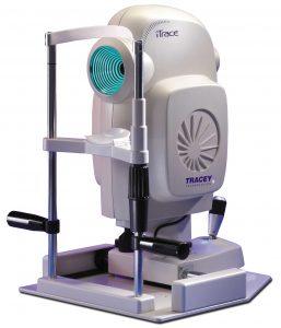
Tracey iTrace Combines autorefraction, corneal topography, auto-keratometry, wavefront aberrometry, and pupillometry in one system. Tracey’s itrace technology enables the trace users to objectively see what the patients see with a total quality visual assessment.
Tracey iTrace also increases the user’s confidence in vision analysis, especially in stressful situations. This is due to its high accuracy and state-of-the-art diagnosis information that improves efficiency while reducing mistakes.
TRACEY iTrace can increase customers’ confidence with accurate information, even in cases of eyesight investigation, enhancing efficiencies, saving time, and avoiding mistakes.
7. Reichert ocular response analyzer (ora) g3 ($11,250.00)

Reichert ocular response analyzer (ora) g3 is the only tonometer that measures corneal hysteresis, a superior predictor of glaucoma progression.
Hysteresis of the corneal is an indication of the biochemical properties. It shows how the cornea differs from the topography, which is the geometrical attribute.
The Ocular Response Analyzer G3 (ORA) is a revolutionary instrument designed to assess the eye’s blood pressure and the retina’s biomechanical properties in an easy, fast measurement.
8. Zeiss Atlas 9000 ($6,666.00)
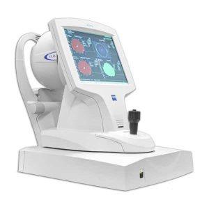
Zeiss Atlas 9000 is a perfect system for the most significant products of corneal data points. It analyzes several images during the alignment and several high-quality photos; it helps streamline the fitting of gas-permeable lenses and guides how to go through difficulty and specialty fittings.
The Zeiss ATLAS 9000 displays corneal higher-order aberrations, providing insight into invaluable treatment planning and education.
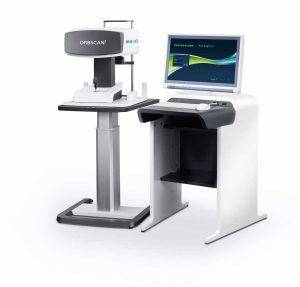
9. OPD Scan III ($9,999.00)
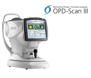
OPD Scan III is a third-generation combined aberrometer and a corneal topographer in the market. The old scan ii is faster, more accurate, and user-friendly than the others initially in the market. This combination, therefore, offers eye care practitioners versatility as a single unit.
Thus, Opd scan iii provides broad information on the eye. This enhances comprehensive analysis and assessment, depending on the model used.
OPD Scan III Wavefront system from Nidek and Marco is a refractive power/corneal analyzer that can evaluate a patient’s overall visual system.
10. Tomey TMS-5 ($15,999.00)

Tomey tms-5 automatically captures several slices by focusing the light alignments on the cornea center in the ring topography. In action, it can take about half to one second, like the measurement time of ring topography.
TOMEY TMS-5 is indicated for cross-sectional imaging of anterior section components of the human eye, like the anterior and anterior surface of the cornea and the anterior chamber, and dimension measurements such as curvature, length, and area by computed analysis.
Is Corneal Topography worth it for your eye clinic?
Because you cannot see or diagnose your eye by yourself, the corneal topography is suitable for the eye clinic. This is because corneal topography will analyze all the changes on the surface of the cornea of your eye. This makes it easy for the doctor to carry out treatment and prescribe drugs to use based on the data points from the optical corneal topography.
An eye is a delicate tissue within living organisms that requires a lot of care and monitoring. However, when I come to eye treatment, the naked eye cannot detect some changes. This means that tests must ensure the procedure is done based on what is present or found occurring in the eye.
Corneal topography helps in making diagnoses and management of the eye. Besides, it also provides the doctor with details that might be required. From the discussion above, it is clear that Corneal Topography Machines are very significant in eye care management.

Dr. Oliver T. Brooks received his undergraduate degree in chemistry from Morehouse College. He received his M.D. degree from Howard University College of Medicine. He completed a residency in Pediatrics at Children’s Hospital- Oakland.
He practiced in the underserved communities of North Oakland and Richmond for four years before accepting a pediatric position at the Watts Health Care Corporation where he is presently Associate Medical Director and Chief of Pediatric and Adolescent Medicine, Chairman of the Quality Management Committee.
