The Keratograph 5M Topographer is a sophisticated corneal topographer that utilizes a built-in real keratometer, along with a color camera optimized for external imaging. Unique features include analyzing the meibomian glands, non-invasive tear film break-up time, and tear meniscus height measurement, as well as assessing the lipid layer.
The OCULUS Keratograph 5M combines corneal topography and iridometric eye evaluation in one machine. The machine utilizes Placido disc illumination to measure the ocular surface, and different colors of light-emitting diodes (LEDs) are employed depending on the application.
The Keratograph 5M features a high-profile color camera that can take both images and video. The apparatus’s built-in software allows me to view 2D and 3D images as needed.
The Oculus Keratograph 5M is a new, patient-friendly, and multi-purpose device that efficiently integrates complex evaluation into a sterile eye examination and generates a comprehensive report. It performs such tests as meibography, eye redness analysis, and Tear Break-Up Time (TBUT). Meibography is an advanced technique that utilizes infrared technology to image the anatomy of the oil glands, located in the upper and lower eyelids. These glands are unique because they provide the natural lubrication to keep the tears wholesome.
It takes place when the oil glands have been compromised (i.e., congested, tortuous, dilated, or atrophied). When assessing MGD, we assess both the anatomy of the glands and the function.
OCULUS Keratograph 5M features:
- Thousands of points used to measure all areas of the cornea, via white ring illumination (Placido Ring Illumination) that also allows for analysis of the tear film to keep glare-related reflex secretion at bay.
- Several illuminations for each function of the Keratograph® 5M, i.e., white diodes for tear-film dynamics, blue diodes for fluo-images, infrared diodes for Meibography.
- This advanced technology can help diagnose dry eye syndrome, enabling the comparison of before-and-after treatment results to assess success.
- A non-invasive test that takes just half an hour to complete, with no touching of the eye!
Dry eye can be a significant problem for patients. In healthy eyes, a fresh layer of tears, called tear film, spreads over the eye when you blink. When this tear film fails to keep the eye moist and comfortable due to insufficient tear production, dry eye syndrome can result.
One benefit of the Keratograph 5M is it is a user-friendly apparatus. The corneal topography portion of the evaluation is non-contact, and the vast majority of the tender eye evaluations are as well — you can use a cotton swab on the eyelid through the meibomian gland topography.
Another significant advantage is that it will help bridge the gap between diagnosing and educating patients about their ocular surface. The system provides me with a simple-to-understand color map and reports that patients can look at and take home together. This enables me to enroll patients in their care and provide them with a personalized treatment plan tailored to their specific ocular needs.
Keratograph 5M Software

JENVIS DRY EYE REPORT
Find the cause of dry eye quickly and reliably. The JENVIS Dry Eye Report is a unique tool for doing this. After measurements are taken using the Keratograph 5M and the slit lamp, your customer/patient receives an easy-to-understand printout. The Dry Eye Report combines screening and consultancy.![]()
TF-SCAN MAKES THE TEAR FILM VISIBLE
Patient consultation made easy. This software shows the quality and quantity of the tear film.
In cases of dry eye patients and contact lens wearers, the tear film should be examined carefully. Only an intact tear film guarantees contact lens wearing comfort! The Keratograph® 4 measures the tear film breakup time non-invasively (quality assessment). You can show your patient the individual tear film quality using the color maps. In addition, you can take another non-invasive measurement to determine the amount of tear film (tear film quantity).
TEAR FILM QUALITY (NIKBUT)
The OCULUS Keratograph® 4 determines the break-up time using the NIKBUT procedure (non-invasive-Keratograph-break-up time).
MENISCUS TEAR HEIGHT ASSESSMENT OF THE TEAR FILM QUANTITY
The height of the torn meniscus can be precisely measured with an integrated ruler and various magnification options, and its development along the edge of the bottom lid can be assessed. The results are saved to the patient file.
LIPID LAYER ASSESSMENT OF THE INTERFERENCE PHENOMENON
The interference colors of the lipid layer and their structure are made visible and can be recorded. The thickness of the lipid layer is assessed based on the structure and color.
TF DYNAMICS ASSESSMENT OF THE PARTICLE FLOW
The video recording, with up to 32 frames per second, enables the observation of the tear film particle flow, from which conclusions regarding the viscosity of the tear film can be drawn.
MEIBO-SCAN MEIBOGRAPHY OF THE TOP AND BOTTOM EYELID
The multi-functionality of the new Keratograph 5M enables even complex examinations, such as Meibography, to be easily and efficiently integrated into routine ophthalmological and optometric check-ups. A dysfunction of the Meibom glands is the most common cause of Dry Eye. The associated morphological changes in the glandular tissue can be made visible with the Meibo-Scan.
PUPILLOMETRY
Indispensable for:
- Fitting multifocal contact lenses
- Exact determination of the treatment area for refractive surgery
- Seamless integration with the existing Keratograph® software
- The infrared camera installed in the Keratograph delivers images of the patient’s pupil, which are used as the basis for the measurements
OPTIONAL IMAGING SOFTWARE*
The Imaging Software is used to record video and image files. In addition to viewing the videos and single images by themselves, you can also compare the recordings with the simulated fluorescein images of the measured eye.
OCULUS OXIMAP®
The OxiMap presents a color map of oxygen transmissibility for soft contact lenses, based on lens power, making it easy to understand – even for your customers.
Include
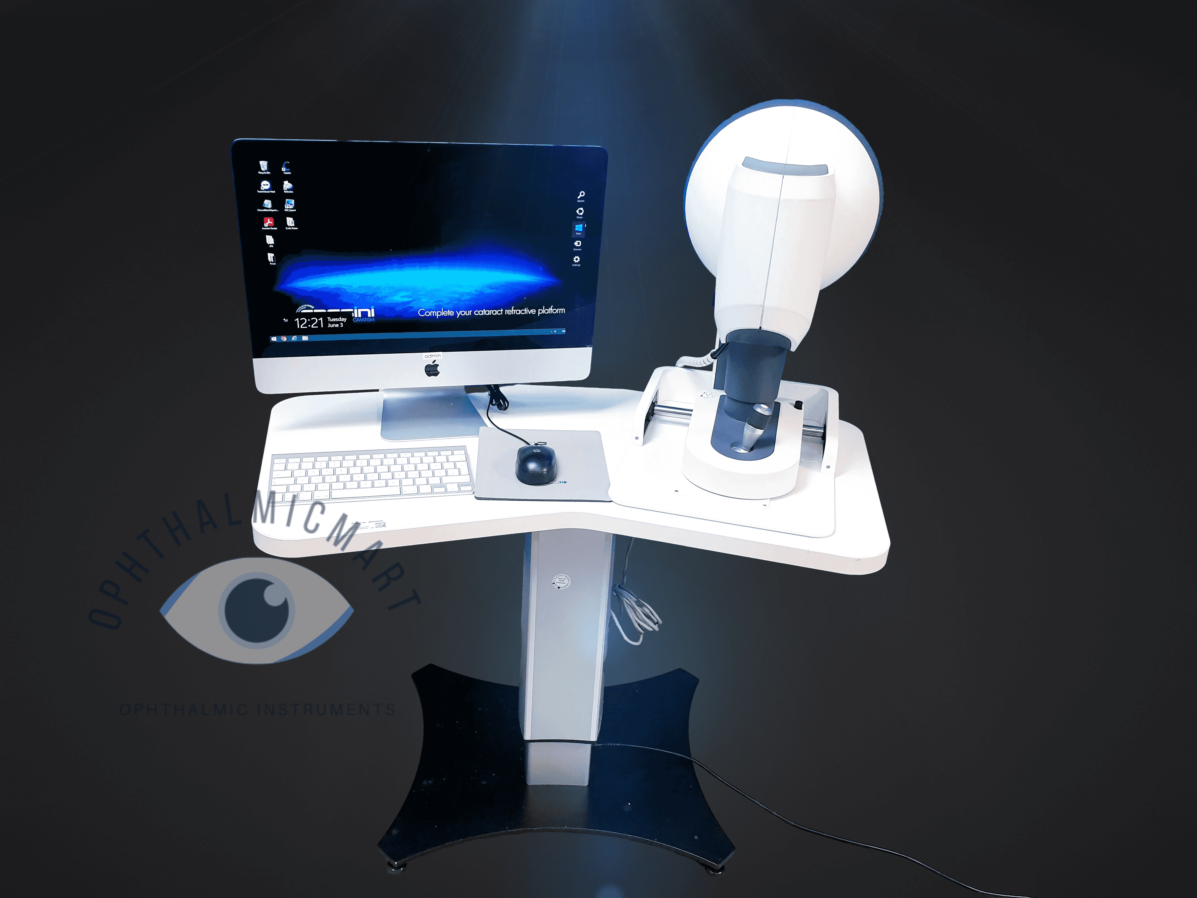
Used, works like new, but no longer needed. Comes with computer/software included, Crystal Report and table if wanted. One of the best all-in-one instruments for dry eye and contact lenses. Includes PC, table, device, installation, training, 1-year warranty, lifetime updates, and online and phone support.