Advancing Smart OCT
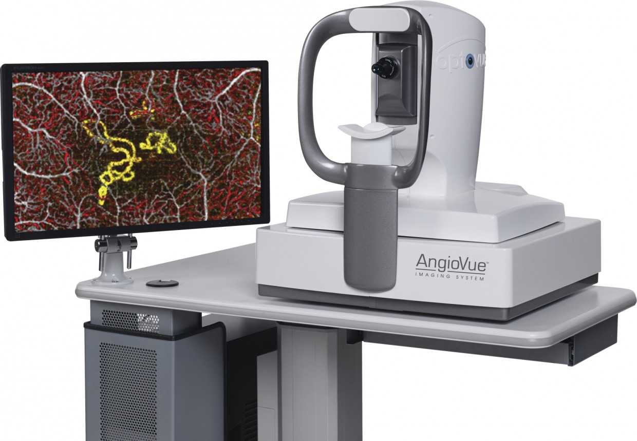
Zeiss Cirrus 5000 HD-OCT is an advanced and innovative smart OCT technology that offers advanced retinal care. With 3D microvascular visualization and industry-leading non-invasive technology, Cirrus HD OCT will improve the quality of services and optimize the efficiency of your practice. It features top of the line visualization, tracking and assessment tools to boost your standard of care.
Zeiss Cirrus 5000 is a clinical assessment tool that features industry defining advancements. It is a quick and effective OCT option which allows you to localize services and improve the level of service provision. These are some few points to note on what the Cirrus HD-OCT has to offer.
What are some features of the Zeiss Cirrus 5000?
Revolutionary assessment options
Up to 6 progression maps are compared during analysis, with areas experiencing statistically significant change being color-coded for easier progression analysis. Alongside the trend analysis of the rate of change, this aspect of assessment will help you collect macular and ONH/RNFL information to address and monitor glaucoma for your patients.
Patient-friendly tracking
Zeiss Cirrus 5000 features Fastrack which is designed to reduce eye motion and focus on the same location throughout multiple visits. The single-pass alignment scanning and unique scan acquisition strategy allow Zeiss Cirrus HD-OCT to focus on an exact spot. You will be able to keep up with your patient’s progress thanks to this tool.
Multiple visualizations for better analysis
Cirrus HD-OCT offers the ability to analyze a single pathology through multiple different views. This ensures comprehensive analysis and insight into a patient’s condition, allowing you to identify and diagnose problems better. You can view cube data from multiple angles thanks to revolutionary 3D imaging, advanced visualization, and OCT fundus images. It also focuses on millions of data points and tightly spaced B-scans to image even the smallest area of pathology.
Why Cirrus HD-OCT will benefit your practice

Comprehensive analysis
The multiple aspects of presentation, including 3D imaging, advanced visualization, and OCT fundus imaging allow for comprehensive analysis of a patient’s condition. You will be better able to examine and monitor your patients, which will allow for more accurate results and improved quality of services.
Ease of operation
The model is easy to use even without a high operational skill level. It is designed for smart scans and en face reports to reduce the workload on your personnel. This allows for better division of labor and efficient delivery of services since it can be operated even by clinicians with minimal technical skill.
3D rendering
The 3D rendering provided by Cirrus HD-OCT allows you to view multiple angles for better assessment. It combines with the auto center function for improved accuracy since this eliminates the need for operator placement of the scope of the examination.
High-speed service provision
Previously captured imaging can be used for comparison, with Cirrus HD-OCT allowing for quick analysis of change. It also features tools such as auto center and Fastrac to enable smooth operation. Patients need only spend a few minutes every time to get comprehensive and accurate results. The high-speed provision of service will improve your efficiency and help to build lasting relationships.
Cirrus HD-OCT for Retinal Disease
With the newest FastTrac retinal monitoring system, exact macular thickness investigations, Fovea Finder, comprehensive layer maps, and more than a hundred B-scans at your disposal, CIRRUS supplies the framework to assess your patient’s retinal condition. CIRRUS data cubes are enrolled with data from previous visits after the scan is obtained. This empowers side-by-side visualization of the exact same location on the retina for every visit. CIRRUS contrasts dimensions from the current and previous visits to extend a thickness change map that helps you determine the next steps to the individual. 3-D cubes and Advanced Visualization™ can be used for pre-operative preparation for VRI disorders. For dry AMD, CIRRUS makes it possible to monitor pre-conversion patients today and prepare for tomorrow’s ironic AMD therapy.