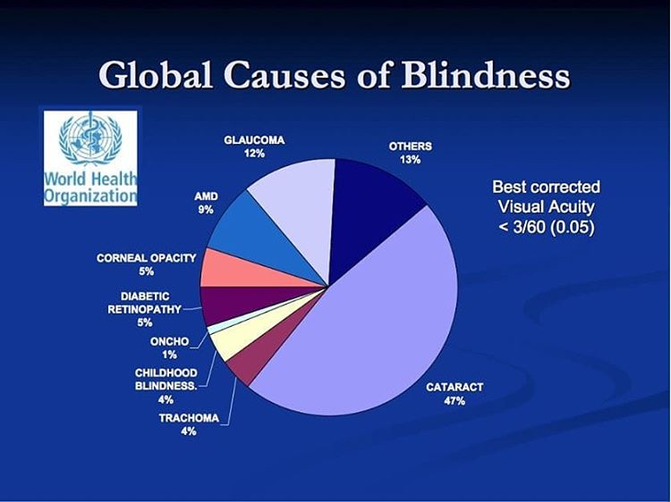
Detecting Glaucoma through regular and complete eye examinations is the secret to protecting your vision from damage. Early detection and careful treatment can maintain vision. Detecting glaucoma in its early phases can be crucial in slowing its progress or preventing vision loss.
Detecting glaucoma early enough can save your eyesight and reverse any complications. How often do you go for eye exams? It would help if you made it a regular habit because eye care is crucial. Glaucoma contributes to high cases of blindness in America. Some people are born with good eyesight and start experiencing blurry vision later in life. If such a person assumes this for long, it could cause glaucoma or a complete loss of sight.
Glaucoma is an eye disorder that damages the optic nerve in the eyes. It comes from accumulating old fluids within the eyes due to a weak drainage system eliminating them. Doctors say you can inherit this disease or even get it from an injury. Some people also develop glaucoma from constant migraines, high blood pressure, or diabetes. Are you above 40 years old? This means that your eyes need more exams compared to a younger person.
A lot of fluids can build pressure on the front area of your eye due to the deterioration of the optic nerve. In normal circumstances, the aqueous humor, a fluid-like substance, flows from the eyes in the form of a mesh-like channel. The blocking of this channel leads to the accumulation of liquids in the eyes, causing glaucoma.
Optometrists know how to diagnose glaucoma by conducting a series of tests. We will discuss some of the diagnostic equipment used by optometrists to diagnose glaucoma. First, look at some criteria that an optometrist can perform on you during an eye exam.
How to detect glaucoma and what Optometrists do.
Tonometry
The first thing an eye doctor does is measure your eye pressure by determining the resistance of your air to the eye. They use a tonometer that can emit an air puff to show the pressure levels of your eye. A pen-like device gets into contact with your eyes to measure the pressure. Your cornea should be clear and shaped regularly for it to function accordingly. Tonometry is used in detecting glaucoma.
Pupil dilation
An optometrist can also dilate your pupils using individual eye drops so that they can view the inside regions of your eyes to determine if everything is in order. They can also conduct a fundus examination to see the inner parts of your eye as they examine the optic nerve and the retina.
Visual field testing
You may also go through this testing that focuses on all the regions of your eyes—the test documents both central and side vision. As the patient, you must press a button every time you see a flash of light during the test.
Pachymetry
An optometrist can also evaluate how thick your cornea is by using an instrument known as an ultrasonic wave. This can also assess your eye pressure. Sometimes thick corneas that are out of the norm portray inaccurate IOP readings. They can either be high or low.
The doctor conducts this test by numbing your eye with an anesthetic drop, then using a probe to emit a painless ultrasound wave. The examination tip has to contact your cornea for the doctor to measure its thickness.
Gonioscopy
Glaucoma comes in different types, and this test can diagnose the closed-angle kind of glaucoma. A doctor can focus on the anterior chamber of your eye using a particular instrument to check if the iris is in the right position. The test can assess the drainage canal between your iris and the cornea. This type of glaucoma is a serious condition that can cause vision loss, thus the need for the doctor to test the drainage canal.
A doctor conducts Gonioscopy using a lens containing a mirror to view the drainage angle found around the edges of the cornea. A doctor puts this lens in your eye after numbing the area using an anesthetic drop. The lens can quickly inform the doctor if the angle is open.
What is the equipment used in detecting glaucoma?
Here is the Top 5 Optometrist diagnostic equipment in diagnosing and detecting glaucoma.
1. Iridex cyclo g6 ($14,999.00) -Safe and effective equipment

Doctors use Iridex Cyclo g6 during transscleral CPC. It is a procedure that comes with numerous benefits for the patient. Iridex Cyclo g6 does not cause inflammation, meaning you don’t have to worry about swollen eyes.
There are also no side effects such as chronic hypotony or phthisis from using this machine. You may not experience any long-term side effects because the device does not produce unnecessary heating.
Product Description
Disposable probes
Iridex Cyclo g6 has various examinations that deliver energy. Professionals know how to diagnose glaucoma using the MP3 investigation, detecting both moderate and mild elevated pressure. They also use the G-probe in the case of elevated eye pressure. They make use of the G-probe with high energy in severe glaucoma cases. The equipment allows doctors to choose the right energy settings for different treatment options. A doctor can, therefore, customize treatment according to a patient’s condition.
Micropulse technology
Iridex Cyclo g6 is equipped with this technology, ensuring tissues can cool down between laser pulses to prevent tissue damage. It, therefore, reduces the risks of treatment to give compelling clinical results. It helps in the treatment of eye diseases such as glaucoma. There is also the use of a legacy laser mechanism that handles detachments and retinal tears. The laser also helps in minimizing intraocular pressure.
Effective procedure
The procedure using this equipment is effective and repeatable. The eye doctor can repeat the process if the initial treatment does not produce the desired results. They can modify the energy levels for repeat treatments using this equipment.
It also offers logistical flexibility since a doctor can perform the procedure from his office or even in an operation room. They can also do it alongside other methods, such as cataract surgery. It is cost-effective, does not include incisions, and is safe to use. The equipment is durable and offers attractive reimbursement for optometrists.
Wide Range of use
The machine can also be used widely on a patient who has never undergone incisional surgery or one who has undergone it. Any patient with a high risk of glaucoma can try to get diagnosed using this equipment.
Key features
- Numerous probes
- Energy settings
- Micropulse technology
2. Nidek RS-3000 ($14,999.00) –High-quality eye photography equipment

Get a machine that comprehensively analyzes your retina and can help detect glaucoma. NIDEK RS-3000 is a photography device that captures accurate images in 3d thanks to its SLO-based eye tracer.
NIDEK RS-3000 can monitor eye movements and help you obtain quality scans that you can use to evaluate the condition of your patient’s eyes.
Product Description
Optical Coherence Tomography sensitivity
This sensitivity allows you to acquire B scan images that are of high resolution through media opacities. You can choose from regular, beautiful, or ultrafine OCT. This technique utilizes invisible light wavelengths to create detailed images from the back region of the eyes.
Physicians, therefore, know how to diagnose glaucoma by using these images, which provide them with the necessary information. They can monitor any changes in your eyes during treatment.
Tracing HD
You can get up to 120 images using the Tracing HD on this equipment. The tracing HD can also display the involuntary movement of the eyes so that you can obtain accurate images. This machine can also detect retinal pathology.
Wide area scan
Using the normative database, it also provides a complete area scan for glaucoma analysis. It also gives you scans at high speed to save time and increase efficiency. A glaucoma patient does not have to wait long for the results when an optometrist uses this equipment. High scanning speed also minimizes artifacts allowing the doctor to get high-quality images.
Key features
- Image averaging: Up to 120 images
- OCT selectivity: Fine, ultra-fine, and regular
- Internal fixation target: laser cross shape
- Normative database: 9×9 mm
3. Humphrey Field Analyzer (HFA) 3 ($29,999.00) –High-speed testing equipment
Every eye doctor should get Humphrey Field Analyzer 3 which can streamline your workflow. The manufacturers equip it with the SIT mechanism that reduces testing time. It utilizes only one trial lens meaning the set-up time is also reduced.
Product Description
It improves confidence
The field analyzer can quickly review a patient’s eye position from different angles to help detect glaucoma. Its effectiveness boosts the determination of a doctor with the test results.
Humphrey Field Analyzer 3 also has a smart touch interface that simplifies operation. To use the interface, you only need to select your patient’s name and press the start section. The user interface is equipped with impressive color graphics. The interface also minimizes the doctor’s steps to follow when conducting a test.
It is user-friendly
The instrument also has reIYE data that enables the doctor to detect testing problems like droopy lids or eye misalignment on the trial lens holder. They can repeat the testing process in such cases to improve the accuracy of the test results. There is also a gaze tracker that helps a doctor ascertain the reliability of the results. The machine is fitted with a graphical user interface, which gives the doctor a 180-degree view, thus making it user-friendly.
Allows quick data transfer
You can transfer legacy data fast from HFA2 to HFA3. Therefore, the machine gives you peace of mind by allowing you to work smoothly as you easily record the patient’s data. It is also fast to reduce patient fatigue and increase their comfort as they undergo different tests.
Mixed GPA
It also allows the free mixing of SITA standards and SITA fast and makes it easy for a doctor to access a patient’s progression analysis, including different SITA texts.
Liquid lens technology
The machine makes use of fluid pressure to deliver patients with refractive correction. You can load every patient’s refractive correction automatically from a previous test. The technology also makes the setup simple to save you time. It also uses an automated trial lens correction to minimize errors and promote accuracy. The trial lens ensures that the patient has a clear view during testing.
Key features
- Gaze tracker
- ReIYE data
- Trial lens correction
- 180-degree view
- Kinetic perimetry
4. Heidelberg HRT 3 ($2,222.00) –High-resolution 3d images machine

Heidelberg HRT 3 is a laser scanning system that can capture 3d images. Doctors are familiar with diagnosing glaucoma thanks to this machine, which allows one to analyze the pictures of the posterior region of the eyes.
Heidelberg HRT 3 conducts retinal topography and monitors any topographical fluctuations to help detect glaucoma. It can also identify other pre-metric diseases thanks to its tracking mechanism.
Product Description
Versatility
Heidelberg HRT 3 offers many functions to optometrists. For instance, it can help you analyze the edema and slits of the macula and any imperfection on the nerve fibers. A doctor can describe the optic nerve head of a glaucoma patient with the help of this machine. You can also evaluate related anatomical features using the equipment.
Optic disc analysis
Though you may be an expert in the field, optical disc analysis can outperform your interpretation and give accurate test results. Optic disc analysis can detect glaucoma even in patients who don’t get early symptoms of the disease. By focusing on the baseline readings alone, it can recognize superior defects that later develop into glaucoma. The machine also uses a micron resolution that provides an in-depth analysis of the corneal structure.
Key features
- Selectable databases
- Network ready
- Asymmetry analysis
5. Tomey casia 2 ($4,600.00) –A user-friendly and fast machine

One of the most outstanding tools created to assist eye doctors in functioning is Tomey cassia 2. It fulfills the needs of a different specialist in eye care treatment since it is fast and accurate. The measurement speed of this machine is unmatched in helping you, from analyzing patients’ data to creating the final report.
Product Description
Advanced technology
The technology used to make Tomey Casia 2 facilitates deep imaging for you to get more information on the different regions of the cornea. Tomey Casia 2 is highly sensitive to depth to capture images that may not be clear using other machines.
Tomey Casia 2 allows you to conduct corneal shape analysis as you analyze different angles to check for any eye complications in a patient. Advanced technology allows you to run tests without necessarily using measuring modes. It has a scanning depth of 13 mm and provides high-resolution images. The scanning speed is also high due to the use of advanced technology.
Versatility
Doctors use Tomey Casia 2 to test for eye complications such as cataracts and glaucoma. The device can support cataract surgery. It is, therefore, versatile in its function, thus saving you the costs of getting more than one machine to use for eye exams. Unlike other tools, this can capture the corneal shape and lens images.
Safety
A doctor can take measurements without bringing the machine in contact with the eye of the patient. It allows non-invasive testing, which reduces the chances of getting an infection from close contact. This makes it safe to use among many patients.
Key features
- Scan depth: 13mm
- Scanning speed: 50.000 A-Scan/sec
- Automated handling
Conclusion
Our eyes may appear similar, but they are very different. Color is not the only differentiating factor in the eyes. Normal eye pressure is also unusual among individuals. Monitoring your eye pressure can help you detect any diseases such as glaucoma.
This disease does not notify anyone of its presence. It comes without any apparent symptoms and keeps progressing without a patient’s knowledge.
Detecting glaucoma through undergoing frequent eye examinations can be a lifesaver. An optometrist can help you assess eye pressure and evaluate optic nerve health. You should also have some necessary information regarding the overall health of your parents because diseases such as glaucoma can be hereditary. Watch for other health conditions, such as hypertension and diabetes, as you maintain a healthy lifestyle.
Statistics reveal that at least 2% of the American population has glaucoma. It mostly affects people above 35 years and those with other medical conditions. Some eye conditions may be hard to treat; therefore, call for a second view from an eye specialist. Get one of the diagnostic equipment reviewed above to ensure accurate results.
Eye screening is how to diagnose glaucoma early enough to prevent vision loss. You may not have vision symptoms at first, but this should not be an excuse to skip an eye exam. A dilated eye exam focuses on the back of your eyes to check for any subtle changes in your optic nerve. It would be best if you went to these tests once or twice a year. It is always better to be safe than sorry.

Dr. Oliver T. Brooks received his undergraduate degree in chemistry from Morehouse College. He received his M.D. degree from Howard University College of Medicine. He completed a residency in Pediatrics at Children’s Hospital- Oakland.
He practiced in the underserved communities of North Oakland and Richmond for four years before accepting a pediatric position at the Watts Health Care Corporation where he is presently Associate Medical Director and Chief of Pediatric and Adolescent Medicine, Chairman of the Quality Management Committee.

One thought on “Optometrist diagnostic equipment in the diagnosis and detecting glaucoma”